| Cite this article as: |
Mohammad Khodaei, Kamran Amini, Alireza Valanezhad, and Ikuya Watanabe, Surface treatment of titanium dental implant with H2O2 solution, Int. J. Miner. Metall. Mater., 27(2020), No. 9, pp. 1281-1286. https://doi.org/10.1007/s12613-020-2016-1

|
Alireza Valanezhad E-mail: vala@nagasaki-u.ac.jp
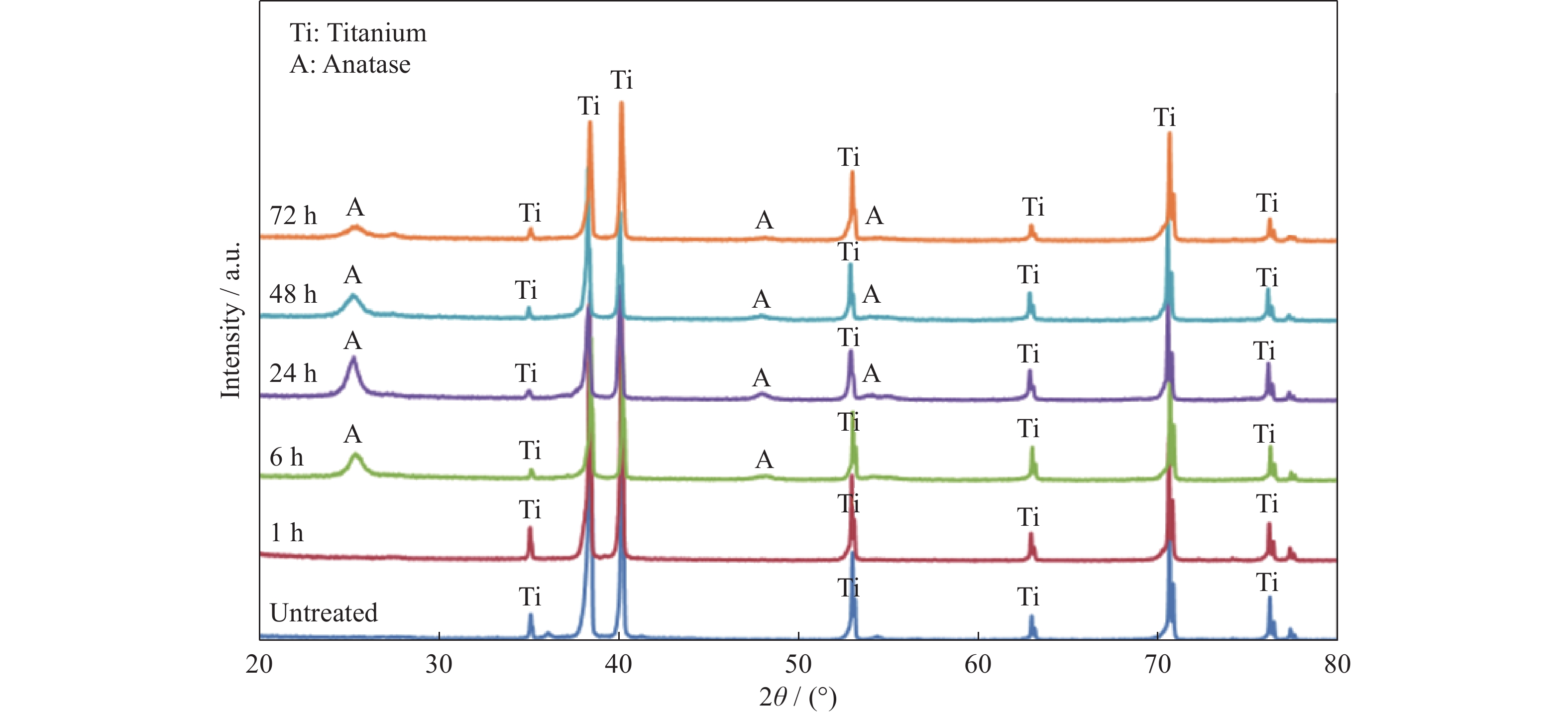
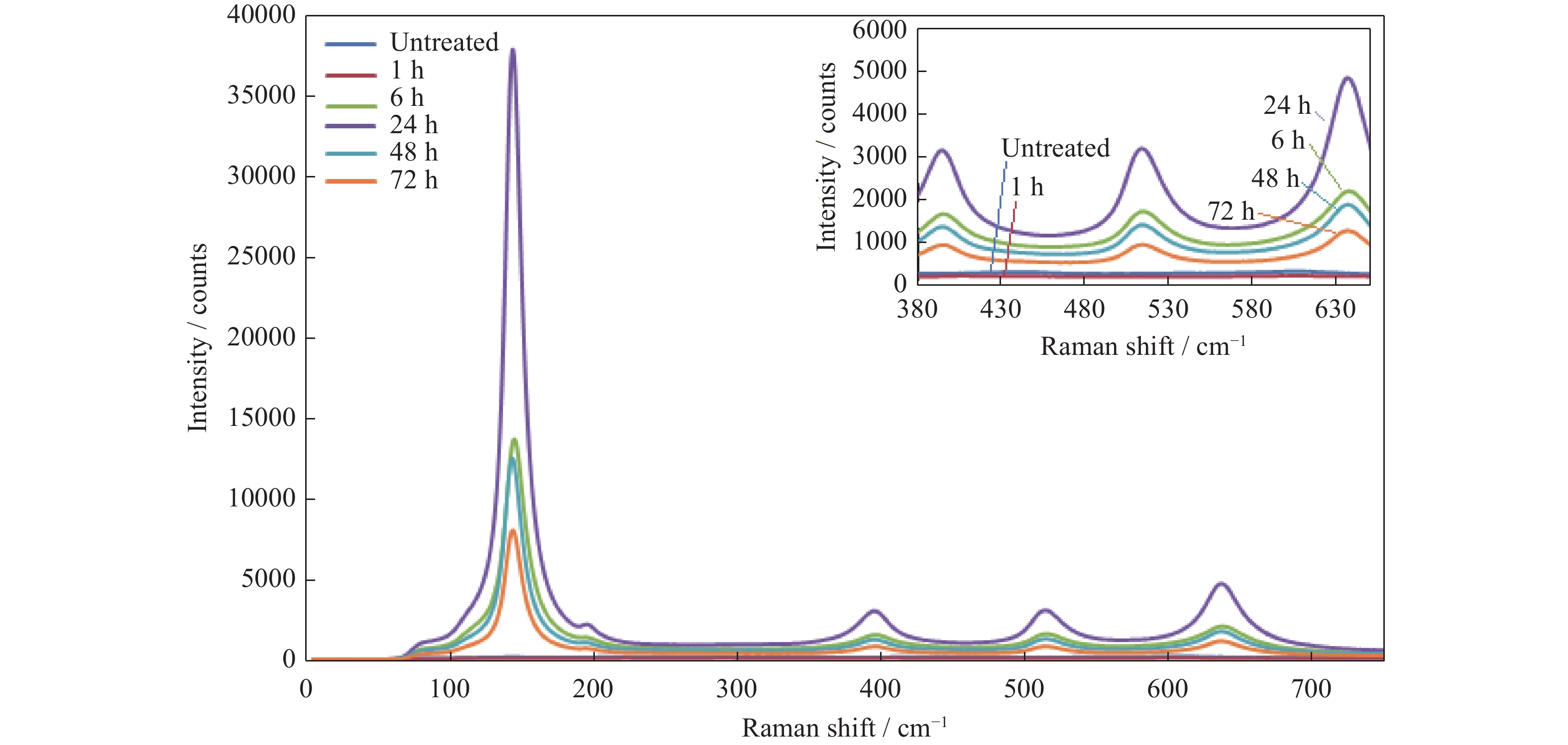
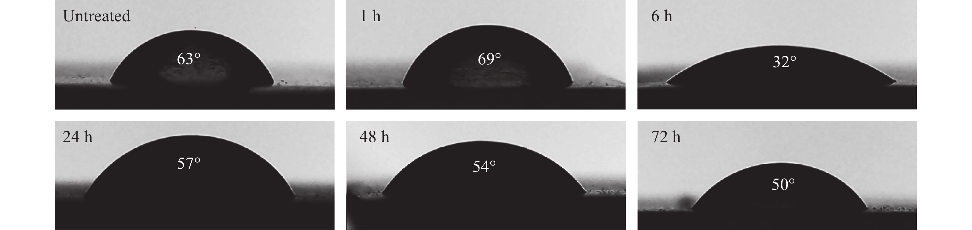
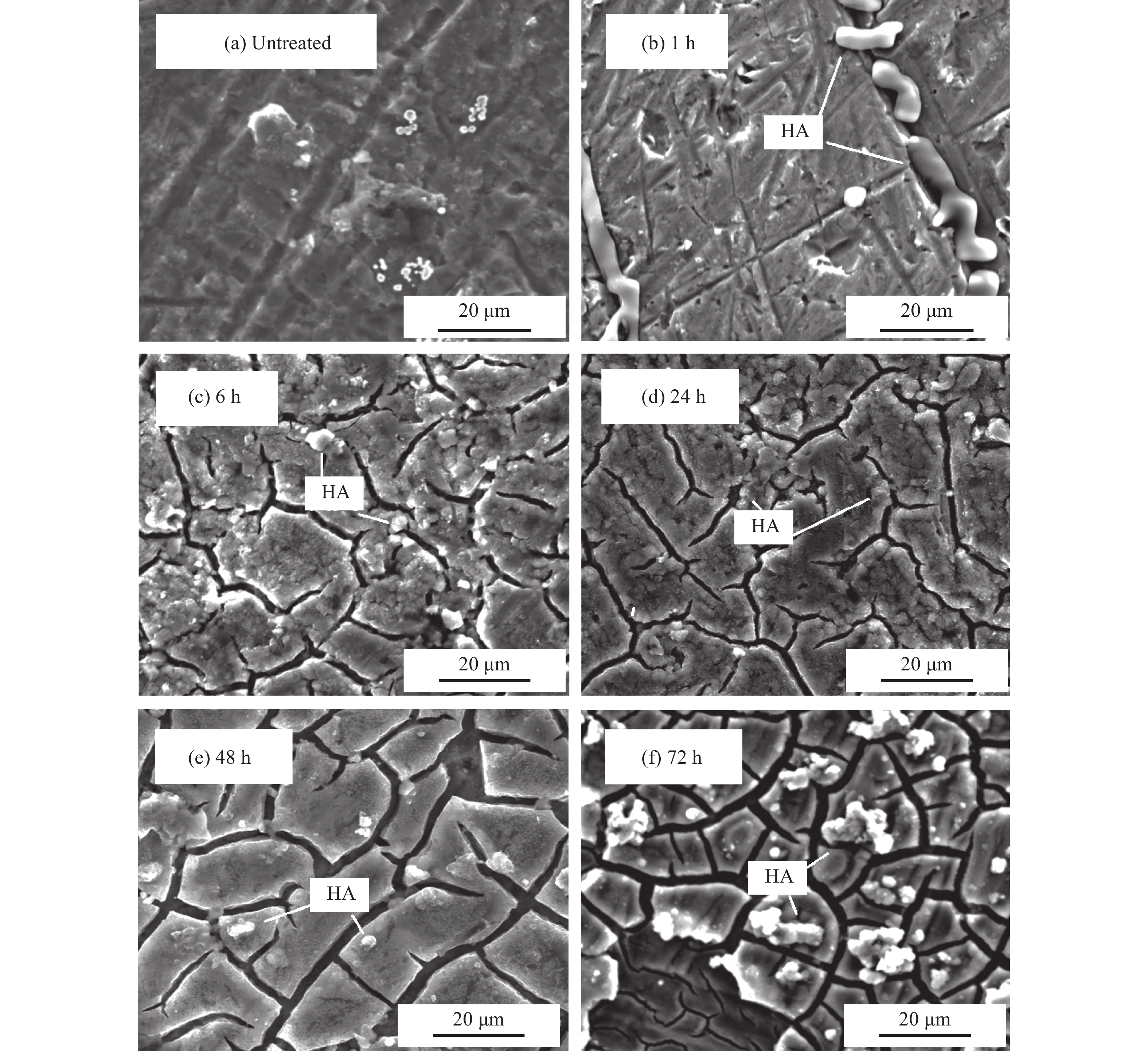
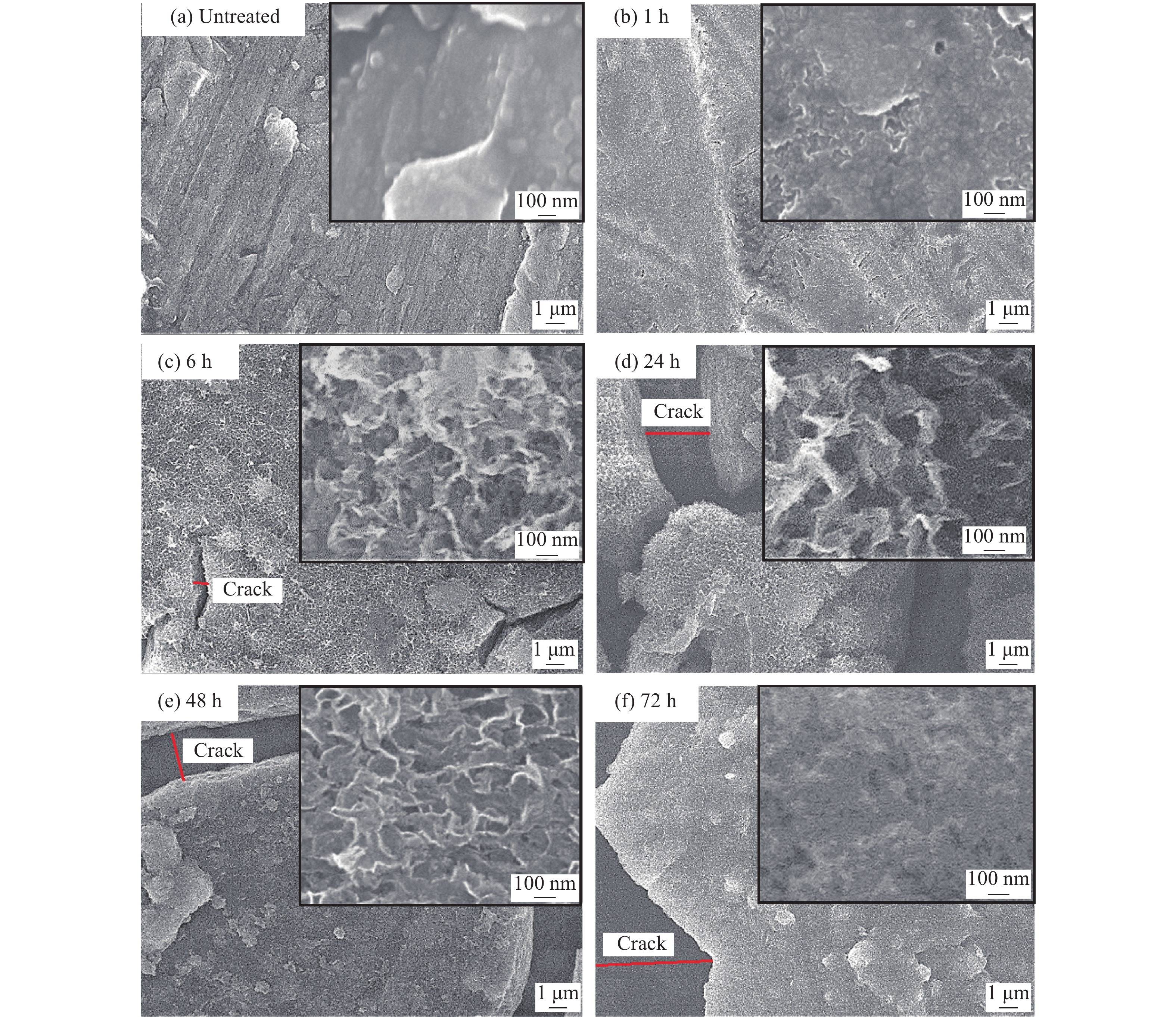
The surface treatment is important for titanium and its alloys as promising candidates for dental implantation due to their bioinert surface. Titanium surface samples were modified using H2O2 solution at different times up to 72 h to boost their bioactivity. According to the results of the field emission scanning electron microscopy test, some nanostructures are formed on the surface of treated titanium samples and increased in size by increasing the time of treatment up to 24 h. After 24 h of application, the sharpness of nanostructures decreased and the micro-cracks and discontinuity in the coating surface increased. The results of the X-ray diffraction study and Raman spectroscopy revealed that anatase (TiO2) was formed on the surface of treated titanium samples. The peak intensity of Raman spectroscopy increased with an improvement in treatment time of up to 24 h and then decreased due to the discontinuity of the coating. Full wettability and ability to form apatite were reached at 6 h of treatment. It is clear that the treatment time has a significant effect on the surface treatment of titanium using the H2O2 solution.

| [1] |
M. Kaur and K. Singh, Review on titanium and titanium based alloys as biomaterials for orthopaedic applications, Mater. Sci. Eng. C, 102(2019), p. 844. doi: 10.1016/j.msec.2019.04.064
|
| [2] |
J.M. Wu, M. Wang, Y.W. Li, F.D. Zhao, X.J. Ding, and A. Osaka, Crystallization of amorphous titania gel by hot water aging and induction of in vitro apatite formation by crystallized titania, Surf. Coat. Technol., 201(2006), No. 3-4, p. 755. doi: 10.1016/j.surfcoat.2005.12.025
|
| [3] |
A. Pandit, J. Planell, and M. Navarro, Titanium and Nitinol (NiTi), 3rd ed., Elsevier, Amsterdam, 2013, p. 120.
|
| [4] |
R.A. Zavanelli, G.E.P. Henriques, I. Ferreira, and J.M. de Almeida Rollo, Corrosion-fatigue life of commercially pure titanium and Ti–6Al–4V alloys in different storage environments, J. Prosthet. Dent., 84(2000), No. 3, p. 274. doi: 10.1067/mpr.2000.108758
|
| [5] |
M. Wei, H.M. Kim, T. Kokubo, and J.H. Evans, Optimising the bioactivity of alkaline-treated titanium alloy, Mater. Sci. Eng. C, 20(2002), No. 1-2, p. 125. doi: 10.1016/S0928-4931(02)00022-X
|
| [6] |
J.M. Wu, Low-temperature preparation of titania nanorods through direct oxidation of titanium with hydrogen peroxide, J. Cryst. Growth, 269(2004), No. 2-4, p. 347. doi: 10.1016/j.jcrysgro.2004.05.023
|
| [7] |
M. Khodaei, A. Valanezhad, I. Watanabe, and R. Yousefi, Surface and mechanical properties of modified porous titanium scaffold, Surf. Coat. Technol., 315(2017), p. 61. doi: 10.1016/j.surfcoat.2017.02.032
|
| [8] |
M. Kosmulski, The significance of the difference in the point of zero charge between rutile and anatase, Adv. Colloid Interface Sci., 99(2002), No. 3, p. 255. doi: 10.1016/S0001-8686(02)00080-5
|
| [9] |
V.M. Frauchiger, Anodic Plasma-chemical Treatment of Titanium Implant Surfaces [Dissertation], Eidgenossische Technische Hochschule Zürich, Zurich, 2002.
|
| [10] |
D.J. Kim, S.H. Hahn, S.H. Oh, and E.J. Kim, Influence of calcination temperature on structural and optical properties of TiO2 thin films prepared by sol–gel dip coating, Mater. Lett., 57(2002), No. 2, p. 355. doi: 10.1016/S0167-577X(02)00790-5
|
| [11] |
M. Khodaei, M. Meratian, O. Savabi, M.H. Fathi, and H. Ghomi, The side effects of surface modification of porous titanium implant using hydrogen peroxide: Mechanical properties aspects, Mater. Lett., 178(2016), p. 201. doi: 10.1016/j.matlet.2016.04.210
|
| [12] |
M. Khodaei, M. Meratian, M. Shaltooki, B. Hashemibeni, O. Savabi, and M. Razavi, Surface modification of Ti6Al4V implants by heat, H2O2 and alkali treatments, Surf. Eng., 32(2016), No. 10, p. 786. doi: 10.1080/02670844.2016.1159818
|
| [13] |
M. Karthega, S. Nagarajan, and N. Rajendran, In vitro studies of hydrogen peroxide treated titanium for biomedical applications, Electrochim. Acta, 55(2010), No. 6, p. 2201. doi: 10.1016/j.electacta.2009.11.057
|
| [14] |
M. Wen, J.F. Gu, G.Liu, Z.B. Wang, and J. Lu, Surface evolution of a gradient structured Ti in hydrogen peroxide solution, Appl. Surf. Sci., 254(2008), No. 9, p. 2905. doi: 10.1016/j.apsusc.2007.10.035
|
| [15] |
M. Khodaei and S.H. Kelishadi, The effect of different oxidizing ions on hydrogen peroxide treatment of titanium dental implant, Surf. Coat. Technol., 353(2018), p. 158. doi: 10.1016/j.surfcoat.2018.08.037
|
| [16] |
T. Kokubo and H. Takadama, How useful is SBF in predicting in vivo bone bioactivity?, Biomaterials, 27(2006), No. 15, p. 2907. doi: 10.1016/j.biomaterials.2006.01.017
|
| [17] |
A. Rodriguez-Contreras, D.G. Bello, and A. Nanci, Surface nanoporosity has a greater influence on osteogenic and bacterial cell adhesion than crystallinity and wettability, Appl. Surf. Sci., 445(2018), p. 255. doi: 10.1016/j.apsusc.2018.03.150
|
| [18] |
W.Y. Meng, Y.M. Zhou, Y.J. Zhang, Q. Cai, L.M. Yang, J.H. Zhao, and C.Y. Li, Osteoblast behavior on hierarchical micro-/nano-structured titanium surface, J. Bionic Eng., 8(2011), No. 3, p. 234. doi: 10.1016/S1672-6529(11)60031-0
|
| [19] |
Q. Liu, W.J. Li, L. Cao, J.J. Wang, Y.M. Qu, X.Y. Wang, R.X. Qiu, X. Di, Z.B. Wang, and B.J. Liang, Response of MG63 osteoblast cells to surface modification of Ti–6Al–4V implant alloy by laser interference lithography, J. Bionic Eng., 14(2017), No. 3, p. 448. doi: 10.1016/S1672-6529(16)60410-9
|
| [20] |
A. Roguska, M. Pisarek, A. Belcarz, L. Marcon, M. Holdynski, M. Andrzejczuk, and M. Janik-Czachor, Improvement of the bio-functional properties of TiO2 nanotubes, Appl. Surf. Sci., 388(2016), p. 775. doi: 10.1016/j.apsusc.2016.03.128
|
| [21] |
P.L. Jiang, J.H. Liang, and C.J. Lin, Construction of micro–nano network structure on titanium surface for improving bioactivity, Appl. Surf. Sci., 280(2013), p. 373. doi: 10.1016/j.apsusc.2013.04.164
|
| [22] |
Y. Luo, S.M. Ge, and Z.M. Jin, Wettability modification for biosurface of titanium alloy by means of sequential carburization, J. Bionic Eng., 6(2009), No. 3, p. 219. doi: 10.1016/S1672-6529(08)60116-X
|
| [23] |
R.A. Gittens, L. Scheideler, F. Rupp, S.L. Hyzy, J. Geis-Gerstorfer, Z. Schwartz, and B.D. Boyan, A review on the wettability of dental implant surfaces Ⅱ: Biological and clinical aspects, Acta Biomater., 10(2014), No. 7, p. 2907. doi: 10.1016/j.actbio.2014.03.032
|
| [24] |
H. Chouirfa, H. Bouloussa, V. Migonney, and C. Falentin-Daudré, Review of titanium surface modification techniques and coatings for antibacterial applications, Acta Biomater., 83(2019), p. 37. doi: 10.1016/j.actbio.2018.10.036
|
| [25] |
A. Osaka, K. Tsuru, and S. Hayakawa, Titania derived from combined chemical and thermal treatments of titanium: In vitro apatite forming ability, Phosphorus Res. Bull., 17(2004), p. 130. doi: 10.3363/prb1992.17.0_130
|
Figures(5)

Copyright © 2019 Editorial Office of International Journal of Minerals, Metallurgy and Materials 京ICP备13030111号
Supported by:
Beijing Renhe Information Technology Co. Ltd
Email:
info@rhhz.net


 Search


 DownLoad: