| Cite this article as: |
Fatma Unal, Faruk Kaya, and Kursat Kazmanli, Synthesis, characterization and radioluminescence properties of erbium-doped yttria phosphors, Int. J. Miner. Metall. Mater., 28(2021), No. 12, pp. 1983-1990. https://doi.org/10.1007/s12613-021-2269-3

|
Fatma Unal E-mail: fatmaunal@hitit.edu.tr
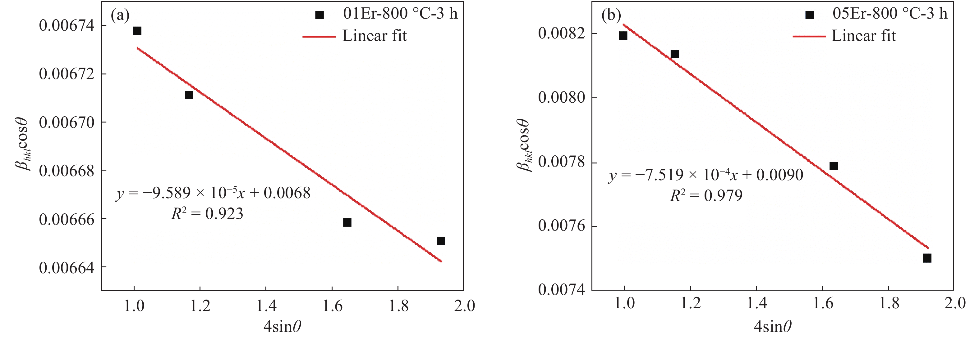
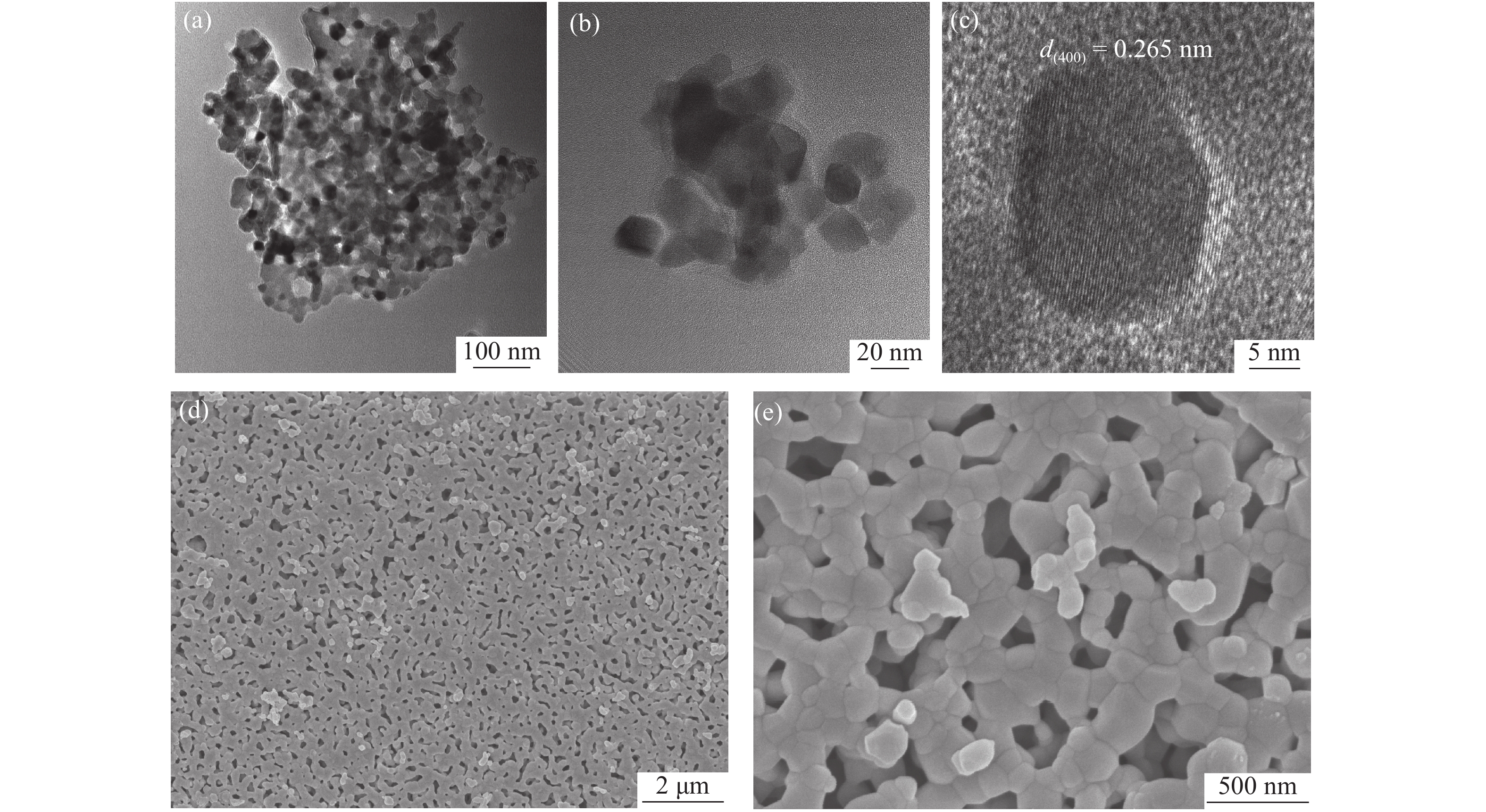
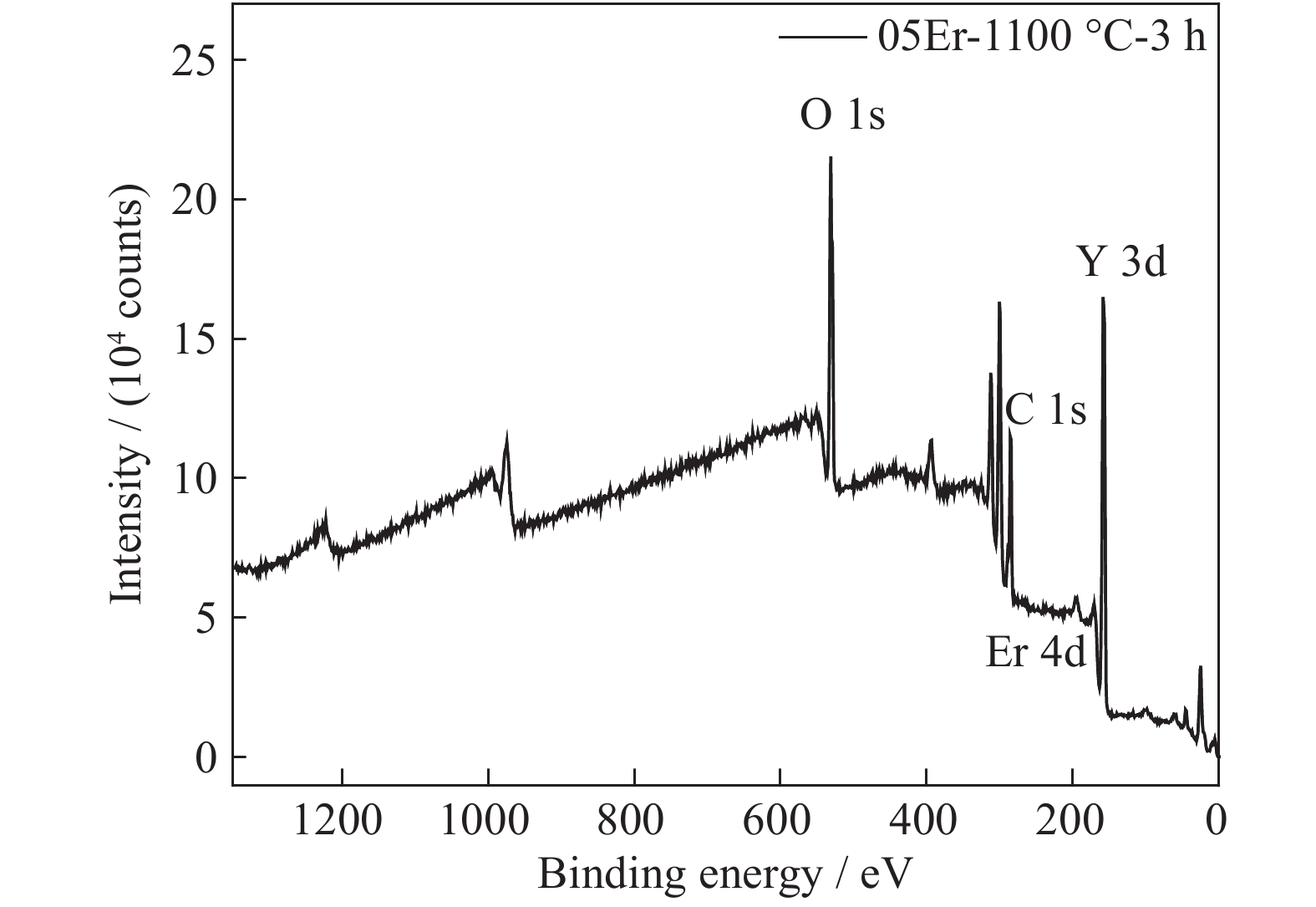
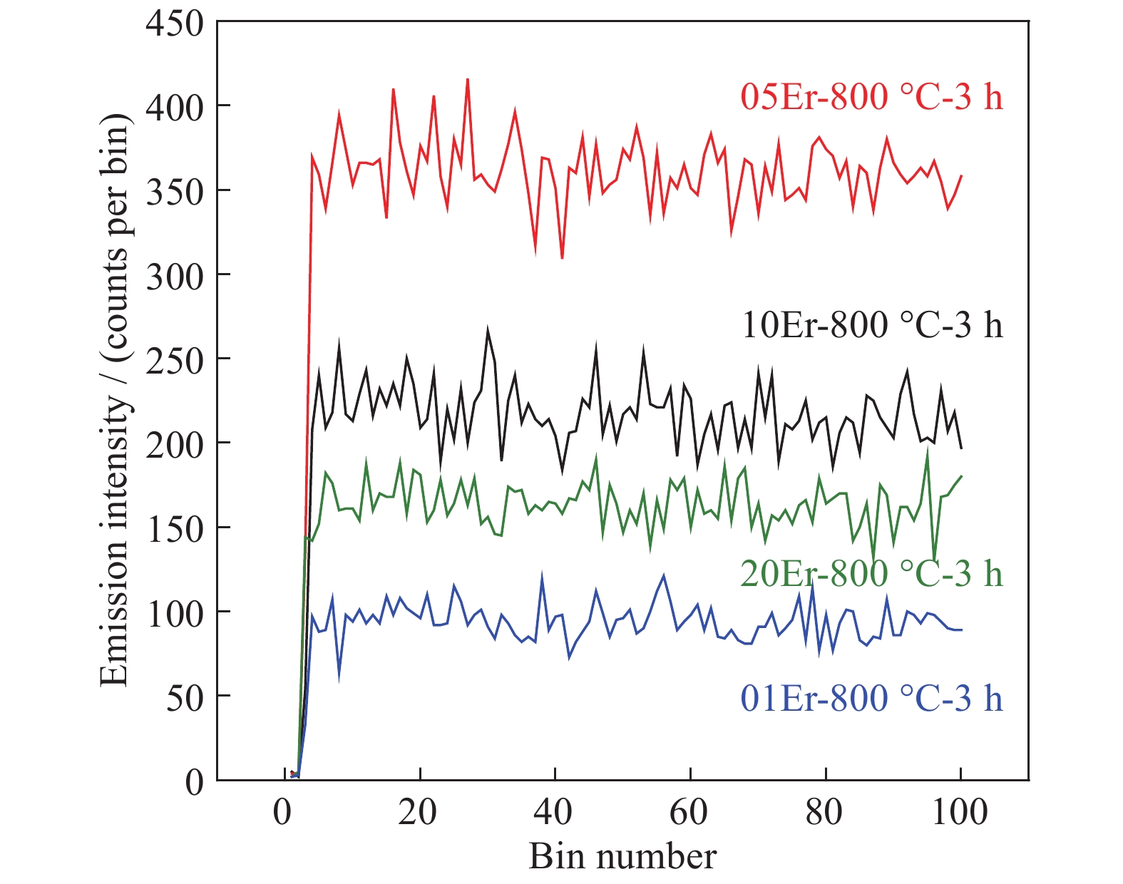
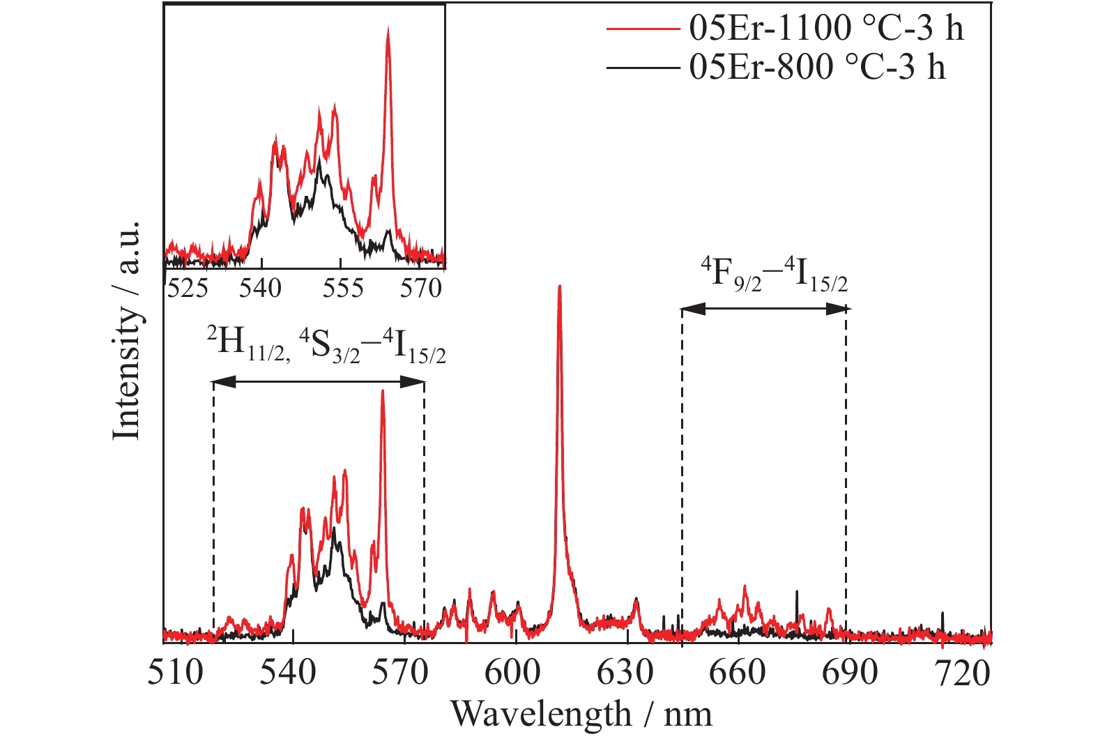
Radioluminescence (RL) behaviour of erbium-doped yttria nanoparticles (Y2O3:Er3+ NPs) which were produced by sol–gel method was reported for future scintillator applications. NPs with dopant rates of 1at%, 5at%, 10at% and 20at% Er were produced and calcined at 800°C, and effect of increased calcination temperature (1100°C) on the RL behaviour was also reported. X-ray diffraction (XRD) results showed that all phosphors had the cubic Y2O3 bixbyite-type structure. The lattice parameters, crystallite sizes (CS), and lattice strain values were calculated by Cohen-Wagner (C-W) and Williamson-Hall (W-H) methods, respectively. Additionally, the optimum solubility value of the Er3+ dopant ion in the Y2O3 host lattice was calculated to be approximately 4at% according to Vegard’s law, which was experimentally obtained from the 5at% Er3+ ion containing solution. Both peak shifts in XRD patterns and X-ray photoelectron spectroscopy (XPS) analyses confirmed that Er3+ dopant ions were successfully incorporated into the Y2O3 host structure. High-resolution transmission electron microscopy (HRTEM) results verified the average CS values and agglomerated NPs morphologies were revealed. Scanning electron microscopy (SEM) results showed the neck formation between the particles due to increased calcination temperature. As a result of the RL measurements under a Cu Kα X-ray radiation (wavelength, λ = 0.154 nm) source with 50 kV and 10 mA beam current, it was determined that the highest RL emission belonged to 5at% Er doped sample. In the RL emission spectrum, the emission peaks were observed in the wavelength ranges of 510–575 nm (2H11/2, 4S3/2–4I15/2; green emission) and 645–690 nm (4F9/2–4I15/2; red emission). The emission peaks at 581, 583, 587, 593, 601, 611 and 632 nm wavelengths were also detected. It was found that both dopant rate and calcination temperature affected the RL emission intensity. The color shifted from red to green with increasing calcination temperature which was attributed to the increased crystallinity and reduced crystal defects.

| [1] |
F. Unal, F. Kaya, and K. Kazmanli, Effects of dopant rate and calcination parameters on photoluminescence emission of Y2O3:Eu3+ phosphors: A statistical approach, Ceram. Int., 45(2019), No. 14, p. 17818. doi: 10.1016/j.ceramint.2019.05.353
|
| [2] |
T. Minami, Y. Kobayashi, T. Miyata, and M. Yamazaki, High-luminance thin-film electroluminescent devices using Y2O3:Mn phosphor, Thin Solid Films, 443(2003), No. 1-2, p. 91. doi: 10.1016/S0040-6090(03)00976-3
|
| [3] |
R.H. Krishna, B.M. Nagabhushana, H. Nagabhushana, N.S. Murthy, S.C. Sharma, C. Shivakumara, and R.P.S. Chakradhar, Effect of calcination temperature on structural. photoluminescence.and thermoluminescence properties of Y2O3:Eu3+ nanophosphor, J. Phys. Chem. C, 117(2013), No. 4, p. 1915. doi: 10.1021/jp309684b
|
| [4] |
L.G. Jacobsohn, B.L. Bennett, R.E. Muenchausen, J.F. Smith, and D.W. Cooke, Optical and structural characterization of nanostructured Y2O3:Tb, [in] Proc. SPIE 6321, Nanophotonic Materials III, San Diego, 2006.
|
| [5] |
R. Pflieger, L. Gravier, G. Guillot, M. Ashokkumar, and S.I. Nikitenko, Inverse effects of the gas feed positioning on sonochemistry and sonoluminescence, Ultrason. Sonochem., 46(2018), p. 10. doi: 10.1016/j.ultsonch.2018.03.019
|
| [6] |
S.S. Pitale, V. Kumar, I.M. Nagpure, O.M. Ntwaeaborwa, E. Coetsee, and H.C. Swart, Cathodoluminescent properties and surface characterization of bluish-white LiAl5O8:Tb phosphor, J. Appl. Phys., 109(2011), No. 1, art. No. 013105. doi: 10.1063/1.3530607
|
| [7] |
F. Unal and K. Kazmanli, Production of un-doped and Er-doped Y2O3 thin films by electron beam evaporation method, Powder Metall. Met. Ceram., 58(2019), No. 3-4, p. 204. doi: 10.1007/s11106-019-00065-0
|
| [8] |
X. Zhang, H.L. Zhao, S. Gao, and Q.F. Zeng, First-principles study of electronic structure and optical properties of Er:Lu2O3, J. Rare Earths, 39(2021), No. 4, p. 453. doi: 10.1016/j.jre.2020.03.001
|
| [9] |
D.A. Permin, S.S. Balabanov, I.L. Snetkov, O.V. Palashov, A.V. Novikova, O.N. Klyusik, and I.V. Ladenkov, Hot pressing of Yb:Sc2O3 laser ceramics with LiF sintering aid, Opt. Mater., 100(2020), art. No. 109701. doi: 10.1016/j.optmat.2020.109701
|
| [10] |
B.P. Gangwar, S. Irusta, and S. Sharma, Physicochemical and optical properties of one-pot combustion synthesized Pr doped La2O3/La(OH)3, J. Lumin., 219(2020), art. No. 116893. doi: 10.1016/j.jlumin.2019.116893
|
| [11] |
Y.Z. Wang, Z.C. Wen, W.G. Ye, Z. Feng, C. Zhao, C.D. Zuo, Y.B. Li, Z.Q. Cao, Z.J. Cao, C.Y. Ma, and Y.G. Cao, Enhanced green up-conversion luminescence in In2O3:Yb3+/Er3+ by tri-doping Zn2+, J. Lumin., 221(2020), art. No. 117029. doi: 10.1016/j.jlumin.2020.117029
|
| [12] |
A. Ćirić and S. Stojadinović, Photoluminescence of Gd2O3 and Gd2O3:Ln3+ (Ln = Eu, Er, Ho) formed by plasma electrolytic oxidation of pure gadolinium substrate, Opt. Mater, 99(2020), art. No. 109546. doi: 10.1016/j.optmat.2019.109546
|
| [13] |
Y.Z. Li, T. Cai, L.X. Yang, S.T. Guo, D. Peng, X.F. Zhao, and Y.Z. Liu, Effect of oxygen partial pressure on the phosphorescence of different lanthanide ion (Ln3+)-doped yttria-stabilised zirconia, Sens. Actuators B, 308(2020), art. No. 127666. doi: 10.1016/j.snb.2020.127666
|
| [14] |
L.X. Yang, D. Peng, X. Shan, F.W. Guo, Y.Z. Liu, X.F. Zhao, and P. Xiao, “Oxygen quenching” in Eu-based thermographic phosphors: Mechanism and potential application in oxygen/pressure sensing, Sens. Actuators B, 254(2018), p. 578. doi: 10.1016/j.snb.2017.07.092
|
| [15] |
T.T. Van, J.R. Bargar, and J.P. Chang, Er coordination in Y2O3 thin films studied by extended x-ray absorption fine structure, J. Appl. Phys., 100(2006), No. 2, art. No. 023115. doi: 10.1063/1.2214299
|
| [16] |
H. Guo and Y.M. Qiao, Preparation, characterization, and strong upconversion of monodisperse Y2O3:Er3+,Yb3+ microspheres, Opt. Mater., 31(2009), No. 4, p. 583. doi: 10.1016/j.optmat.2008.06.011
|
| [17] |
V.K. Rai, A. Pandey, and R. Dey, Photoluminescence study of Y2O3:Er3+–Eu3+–Yb3+ phosphor for lighting and sensing applications, J. Appl. Phys., 113(2013), No. 8, art. No. 083104. doi: 10.1063/1.4793265
|
| [18] |
Q.P. Lu, Y.B. Hou, A.W. Tang, X.J. Liu, and F. Teng, Synthesis of porous Y2O3:Er plates with enhanced upconversion luminescence properties, Mater. Lett., 99(2013), p. 115. doi: 10.1016/j.matlet.2013.02.084
|
| [19] |
T. Hirai, T. Orikoshi, and I. Komasawa, Preparation of Y2O3:Yb,Er infrared-to-visible conversion phosphor fine particles using an emulsion liquid membrane system, Chem. Mater., 14(2002), No. 8, p. 3576. doi: 10.1021/cm0202207
|
| [20] |
T. Nunokawa, O. Odawara, and H. Wada, Optical properties of highly crystalline Y2O3:Er,Yb nanoparticles prepared by laser ablation in water, Mater. Res. Express, 1(2014), No. 3, art. No. 035043. doi: 10.1088/2053-1591/1/3/035043
|
| [21] |
V. Lojpur, L. Mancic, P. Vulic, M.D. Dramicanin, M.E. Rabanal, and O. Milosevic, Structural, morphological and up-converting luminescence characteristics of nanocrystalline Y2O3:Yb/Er powders obtained via spray pyrolysis, Ceram. Int., 40(2014), No. 2, p. 3089. doi: 10.1016/j.ceramint.2013.10.002
|
| [22] |
J.I. Eldridge, Luminescence decay-based Y2O3:Er phosphor thermometry: Temperature sensitivity governed by multiphonon emission with an effective phonon energy transition, J. Lumin., 214(2019), art. No. 116535. doi: 10.1016/j.jlumin.2019.116535
|
| [23] |
C.C. Mi, J.P. Zhang, H.Y. Gao, X.L. Wu, M. Wang, Y.F. Wu, Y.Q. Di, Z.R. Xu, C.B. Mao, and S.K. Xu, Multifunctional nanocomposites of superparamagnetic (Fe3O4) and NIR-responsive rare earth-doped up-conversion fluorescent (NaYF4:Yb,Er) nanoparticles and their applications in biolabeling and fluorescent imaging of cancer cells, Nanoscale, 2(2010), No. 7, p. 1141. doi: 10.1039/c0nr00102c
|
| [24] |
F.L. Meng, Y. Luo, Y.L. Zhou, J.W. Zhang, Y.Z. Zheng, G.Z. Cao, and X. Tao, Integrated plasmonic and upconversion starlike Y2O3:Er/Au@TiO2 composite for enhanced photon harvesting in dye-sensitized solar cells, J. Power Sources, 316(2016), p. 207. doi: 10.1016/j.jpowsour.2016.03.032
|
| [25] |
R. Dey, A. Pandey, and V.K. Rai, Er3+–Yb3+ and Eu3+–Er3+–Yb3+ codoped Y2O3 phosphors as optical heater, Sens. Actuators B, 190(2014), p. 512. doi: 10.1016/j.snb.2013.09.025
|
| [26] |
D.L. Yin, J. Wang, Y. Wang, P. Liu, J. Ma, X.D. Xu, D.Y. Shen, Z.L. Dong, L.B. Kong, and D.Y. Tang, Fabrication of Er:Y2O3 transparent ceramics for 2.7 μm mid-infrared solid-state lasers, J. Eur. Ceram. Soc., 40(2020), No. 2, p. 444. doi: 10.1016/j.jeurceramsoc.2019.09.051
|
| [27] |
T. Yanagida, Study of rare-earth-doped scintillators, Opt. Mater., 35(2013), No. 11, p. 1987. doi: 10.1016/j.optmat.2012.11.002
|
| [28] |
K. Rubešová, T. Thoř, V. Jakeš, D. Mikolášová, J. Maixner, O. Jankovský, J. Cajzl, L. Nádherný, Lanthanide-doped Y2O3 – The photoluminescent and radioluminescent properties of sol–gel prepared samples, Ceram. Silik., 62(2018), No. 4, p. 411.
|
| [29] |
Y. Fujimoto, T. Yanagida, Y. Yokota, A. Ikesue, and A. Yoshikawa, Evaluation of characterization of rare-earth doped sesquioxide ceramic scintillators, Opt. Mater., 34(2011), No. 2, p. 448. doi: 10.1016/j.optmat.2011.03.049
|
| [30] |
I. Kandarakis, D. Cavouras, D. Nikolopoulos, P. Liaparinos, A. Episkopakis, K. Kourkoutas, N. Kalivas, N. Dimitropoulos, I. Sianoudis, C. Nomicos, and G. Panayiotakis, Modelling angular distribution of light emission in granular scintillators used in X-ray imaging detectors, [in] A. Méndez-Vilas, ed., Proceedings of the First International Meeting on Applied Physics (APHYS-2003), Badajoz, 2003, p. 909.
|
| [31] |
N. Kumamoto, D. Nakauchi, T. Kato, G. Okada, N. Kawaguchi, and T. Yanagida, Photoluminescence, scintillation and thermally-stimulated luminescence properties of Tb-doped 12CaO•7Al2O3 single crystals grown by FZ method, J. Rare Earths, 35(2017), No. 10, p. 957. doi: 10.1016/S1002-0721(17)60999-2
|
| [32] |
B.K. Cha, S.J. Lee, P. Muralidharan, J.Y. Kim, D.K. Kim, D.H. Lee, J.I. Yun, and G. Cho, Synthesis and scintillation properties of nano Gd2O3(Eu) scintillator for high resolution X-ray imaging applications, Nucl. Instrum. Methods Phys. Res. Sect. A, 619(2010), No. 1-3, p. 174. doi: 10.1016/j.nima.2009.10.171
|
| [33] |
K. Kamada, K. Hishinuma, S. Kurosawa, A. Yamaji, Y. Shoji, J. Pejchal, Y. Ohashi, Y. Yokota, and A. Yoshikawa, Growth and luminescence properties of Eu-doped HfO2/α-Al2O3 eutectic scintillator, J. Rare Earths, 34(2016), No. 8, p. 796. doi: 10.1016/S1002-0721(16)60096-0
|
| [34] |
S. Fukushima, T. Furukawa, H. Niioka, M. Ichimiya, T. Sannomiya, J. Miyake, M. Ashida, T. Araki, and M. Hashimoto, Synthesis of Y2O3 nanophosphors by homogeneous precipitation method using excessive urea for cathodoluminescence and upconversion luminescence bioimaging, Opt. Mater. Express, 6(2016), No. 3, p. 831. doi: 10.1364/OME.6.000831
|
| [35] |
Y.B. Mao, X. Guo, T. Tran, K.L. Wang, C.K. Shih, and J.P. Chang, Luminescent properties of ensemble and individual erbium-doped yttrium oxide nanotubes, J. Appl. Phys., 105(2009), No. 9, art. No. 094329. doi: 10.1063/1.3117520
|
| [36] |
D. den Engelsen, G.R. Fern, T.G. Ireland, and J. Silver, Cathodoluminescence of Y2O3:Ln3+ (Ln = Tb, Er and Tm) and Y2O3:Bi3+ nanocrystalline particles at 200 keV, RSC Adv., 8(2018), No. 1, p. 396. doi: 10.1039/C7RA12644A
|
| [37] |
D. Avram, B. Cojocaru, I. Tiseanu, M. Florea, and C. Tiseanu, Down-/up-conversion emission enhancement by Li addition : Improved crystallization or local structure distortion?, J. Phys. Chem. C, 121(2017), No. 26, p. 14274. doi: 10.1021/acs.jpcc.7b02897
|
| [38] |
A.S. Hassanien, A.A. Akl, and A.H. Sáaedi, Synthesis, crystallography, microstructure, crystal defects, and morphology of BixZn1−xO nanoparticles prepared by sol–gel technique, CrystEngComm, 20(2018), No. 12, p. 1716. doi: 10.1039/C7CE02173A
|
| [39] |
Z.C. Cordero and C.A. Schuh, Phase strength effects on chemical mixing in extensively deformed alloys, Acta Mater., 82(2015), p. 123. doi: 10.1016/j.actamat.2014.09.009
|
| [40] |
R.V. Perrella, D.P. dos Santos, G.Y. Poirier, M.S. Góes, S.J.L. Ribeiro, M.A. Schiavon, and J.L. Ferrari, Er3+-doped Y2O3 obtained by polymeric precursor: Synthesis, structure and upconversion emission properties, J. Lumin., 149(2014), p. 333. doi: 10.1016/j.jlumin.2014.01.052
|
| [41] |
W. Zhong, H.X. Dai, C.F. Ng, and C.T. Au, A comparison of nanoscale and large-size BaCl2-modified Er2O3 catalysts for the selective oxidation of ethane to ethylene, Appl. Catal. A, 203(2000), No. 2, p. 239. doi: 10.1016/S0926-860X(00)00486-5
|
| [42] |
S. Agrawal and V. Dubey, Down conversion luminescence behavior of Er and Yb doped Y2O3 phosphor, J. Radiat. Res. Appl. Sci., 7(2014), No. 4, p. 601. doi: 10.1016/j.jrras.2014.09.014
|
| [43] |
H. Huang, X.H. Sun, S.D. Wang, Y. Liu, X.R. Li, J.L. Liu, Z.H. Kang, and S.T. Lee, Strong red emission of pure Y2O3 nanoparticles from oxygen related defects, Dalton Trans., 40(2011), No. 43, p. 11362. doi: 10.1039/c1dt11553g
|
| [44] |
The International Commission on Illumination (CIE), Fundamental Chromaticity Diagram with Physiological Axes. Part 1, Technical Report 170-1, CIE Central Bureau, Vienna, 2006.
|

Figures(9) / Tables(3)

Copyright © 2019 Editorial Office of International Journal of Minerals, Metallurgy and Materials 京ICP备13030111号
Supported by:
Beijing Renhe Information Technology Co. Ltd
Email:
info@rhhz.net


 Search


 DownLoad: